What Is a Skin Biopsy and Why Are There Different Types

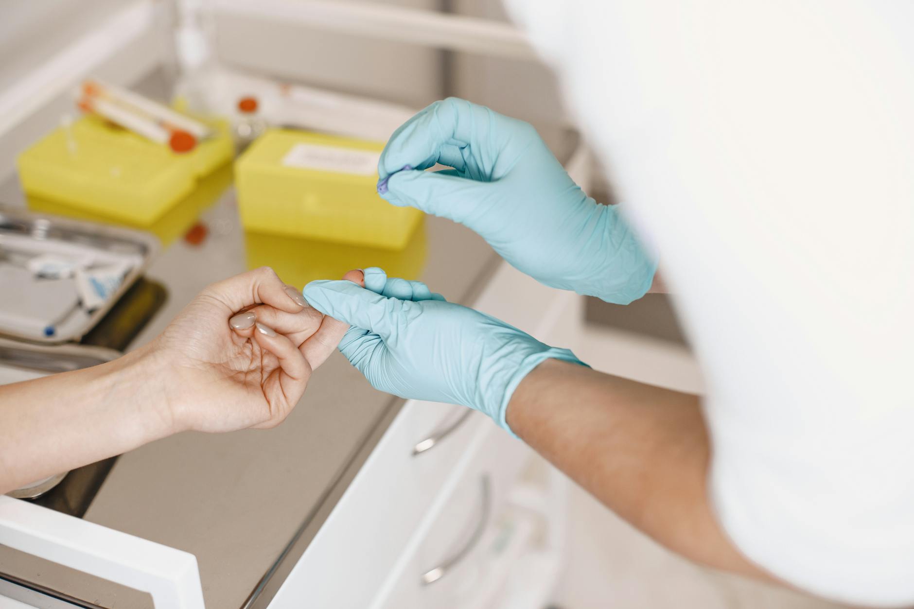
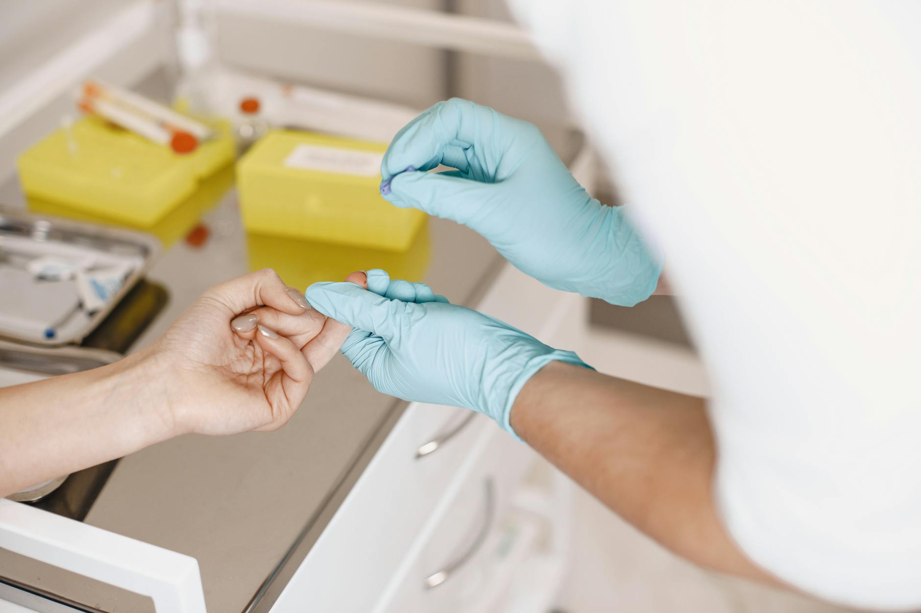
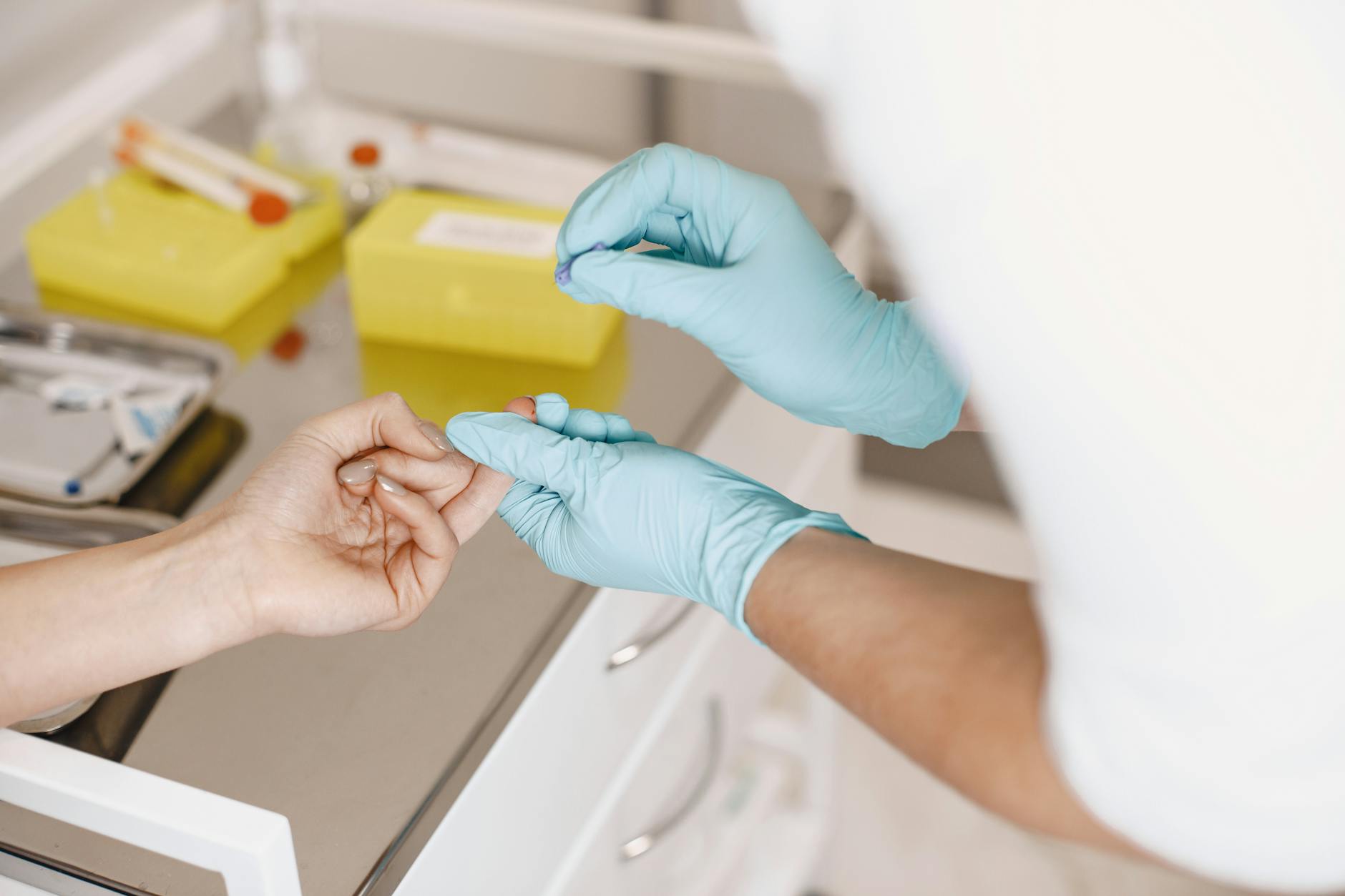
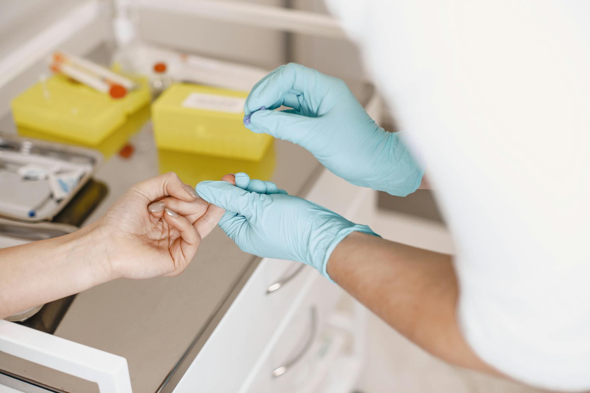
A skin biopsy is a medical procedure where a dermatologist removes a small piece of skin tissue to examine it under a microscope. This diagnostic tool helps doctors identify skin conditions, rule out serious diseases like cancer, and determine the best treatment approach for your specific situation.
“Different biopsy techniques exist because skin conditions vary in depth, location, and the amount of tissue needed for accurate diagnosis.”
Dermatologists use various biopsy methods because your skin has multiple layers, and different conditions affect these layers differently. Some skin problems only involve the surface, while others extend deeper into the tissue. The location of the suspicious area also matters—delicate areas like your face require different approaches than thicker skin on your back.
Understanding that scarring concerns are natural, dermatologists select the least invasive method that still provides enough tissue for diagnosis. Modern biopsy techniques are designed to minimize scarring while ensuring accurate results. Your doctor will choose the specific type based on the size, location, and suspected condition of the area being examined.
Each biopsy method serves a specific purpose, balancing diagnostic accuracy with cosmetic outcomes to address your concerns effectively.
Punch Biopsy: The Most Common Method

A punch biopsy uses a circular blade that looks like a small cookie cutter to remove a cylindrical piece of skin tissue. Your dermatologist rotates this instrument into your skin, creating a clean, round sample that includes all layers from the surface down to the fat beneath. The procedure takes just minutes and provides doctors with comprehensive tissue for accurate diagnosis.
“Punch biopsies create predictably small scars that heal well with proper care, addressing most patients' concerns about permanent marks.”
Dermatologists choose punch biopsies for suspicious moles, rashes, and inflammatory skin conditions where they need to examine the full thickness of skin. The standard sizes range from 3mm to 8mm in diameter, with most procedures using 4mm punches. This method works particularly well for lesions on arms, legs, and torso where slightly larger scars blend naturally with surrounding skin.
The sample goes deep enough to capture important diagnostic information while remaining small enough to close with one or two stitches. For example, when examining a changing mole for melanoma, the punch biopsy provides pathologists with enough tissue to determine cancer type and depth of invasion. Most punch biopsy sites heal within two weeks, leaving a small linear scar that fades significantly over time.
Shave Biopsy: For Surface-Level Concerns
A shave biopsy removes only the top layers of skin using a razor-like blade or scalpel. Your dermatologist numbs the area with local anesthesia, then carefully shaves off the suspicious tissue parallel to the skin surface. This creates a flat, shallow wound that resembles a scrape rather than a deep cut.
This technique works best for surface-level skin concerns like suspected basal cell carcinomas, seborrheic keratoses, or raised moles. Since shave biopsies only remove superficial tissue, they're not suitable for deeper lesions or when your doctor suspects melanoma, which requires examining tissue at various depths.
Recovery involves keeping the wound clean and dry while it heals from the bottom up. Most patients experience minimal scarring since the procedure doesn't penetrate deeply into the skin. You might notice a slightly lighter or darker spot where the biopsy was taken, but this usually fades over several months.
Incisional and Excisional Biopsies: For Larger Areas
When dermatologists need to examine deeper tissue layers or larger suspicious areas, they turn to surgical biopsy methods. These procedures provide more comprehensive tissue samples than surface-level techniques, giving doctors the detailed information needed for accurate diagnosis and treatment planning.
Incisional biopsy removes only a portion of the suspicious tissue, typically used when the lesion is too large to remove entirely or when sampling is sufficient for diagnosis. Your doctor makes a small incision and takes a representative section, leaving the remainder intact. This approach works well for large moles or when testing is needed before deciding on full removal.
Excisional biopsy removes the entire suspicious area along with a margin of healthy tissue around it. This method serves both diagnostic and therapeutic purposes, as it can completely eliminate the problem while providing tissue for analysis. For example, a suspicious mole measuring two centimeters might be entirely removed with surrounding healthy skin to ensure clean margins.
Both procedures require local anesthesia and result in small, neat scars that fade significantly over time. Your dermatologist will discuss which method best suits your specific situation, considering factors like lesion size, location, and suspected diagnosis.
How Your Dermatologist Chooses the Right Type

Your dermatologist considers several key factors when selecting the most appropriate biopsy method for your specific situation. The size and appearance of the suspicious area plays a major role in this decision-making process.
Location matters significantly. Areas with cosmetic importance, like your face, often receive punch biopsies to minimize scarring concerns. Meanwhile, locations with thicker skin, such as your back or shoulders, can accommodate larger excisional procedures without noticeable marks.
The suspected condition strongly influences the choice. If melanoma is suspected, your dermatologist will likely recommend excisional biopsy to examine the entire lesion and determine precise depth. For routine mole checks or inflammatory conditions, a simple shave or punch biopsy provides adequate tissue for diagnosis.
Your dermatologist also considers your medical history, current medications, and healing capacity. Patients taking blood thinners might receive smaller biopsies to reduce bleeding risks. The goal is always obtaining sufficient tissue for accurate diagnosis while minimizing discomfort and scarring potential.
What Happens After Your Biopsy
Your biopsy sample travels to a specialized laboratory where pathologists examine the tissue under microscopes. This process typically takes 7-14 business days, though some cases requiring additional testing may take longer. The pathologist analyzes cellular structures, looking for signs of cancer, inflammation, or other skin conditions.
Your dermatologist will contact you once results arrive, either by phone or through a follow-up appointment. Normal results often come via phone call, while abnormal findings usually require an in-person discussion. Understanding your results doesn't need to cause anxiety about permanent scarring—most skin conditions are highly treatable when caught early.
Follow-up care depends on your specific results. Benign findings may only require routine monitoring, while precancerous or cancerous tissue often needs additional treatment. Some patients benefit from complementary procedures like Chemical Peels, which use controlled chemical exfoliation to improve skin texture and address sun damage that contributed to their original concern.
Proper wound care at your biopsy site promotes healing without scarring. Keep the area clean and dry, follow your dermatologist's specific instructions, and avoid picking at any scabs that form.
Most biopsy results lead to successful treatment outcomes when patients follow their dermatologist's recommendations and maintain proper follow-up care.
Frequently Asked Questions
What is a skin biopsy and why do I need one?
A skin biopsy is a medical procedure where a dermatologist removes a small piece of skin tissue to examine it under a microscope. This diagnostic tool helps doctors identify skin conditions, rule out serious diseases like cancer, and determine the best treatment approach for your specific situation. Different biopsy techniques exist because skin conditions vary in depth, location, and the amount of tissue needed for accurate diagnosis.
What is the difference between a punch biopsy and a shave biopsy?
A punch biopsy uses a circular blade to remove a cylindrical piece of skin tissue that includes all layers from the surface down to the fat beneath, making it suitable for suspicious moles and deeper lesions. A shave biopsy removes only the top layers of skin using a razor-like blade, creating a flat, shallow wound that works best for surface-level concerns like basal cell carcinomas. Punch biopsies are used when doctors need to examine the full thickness of skin, while shave biopsies are limited to superficial tissue.
Will a skin biopsy leave a scar?
Modern biopsy techniques are designed to minimize scarring while ensuring accurate results, and dermatologists select the least invasive method that still provides enough tissue for diagnosis. Punch biopsies create predictably small scars that heal well with proper care, typically leaving a small linear scar that fades significantly over time. Shave biopsies have a 95% minimal scarring rate and heal within 7-14 days, resembling a scrape rather than a deep cut.
How long does it take for a skin biopsy to heal?
Most punch biopsy sites heal within two weeks, leaving a small linear scar that fades significantly over time. Shave biopsies typically have a healing time of 7-14 days and create minimal scarring. The exact healing time depends on the type of biopsy performed, the location on your body, and how well you care for the wound during recovery.
When would a doctor choose a punch biopsy over a shave biopsy?
Dermatologists choose punch biopsies for suspicious moles, rashes, and inflammatory skin conditions where they need to examine the full thickness of skin, particularly when melanoma is suspected. Shave biopsies are not suitable for deeper lesions or when doctors suspect melanoma, which requires examining tissue at various depths. The choice depends on the size, location, and suspected condition of the area being examined, with doctors balancing diagnostic accuracy and cosmetic outcomes.
Disclaimer: This content is for informational purposes only and does not constitute medical advice. Always consult a qualified healthcare provider before making health decisions.